Recently, the ultra-sensitive magnetic resonance team from the Innovation Academy for Precision Measurement Science and Technology (APM) has proposed a novel method for dynamic ventilation function imaging of the lungs with high spatiotemporal resolution. This method has achieved the highest current spatiotemporal resolution for in vivo dynamic ventilation function imaging of the lungs, with a spatial resolution reaching the sub-millimeter level (0.5 mm) and a temporal resolution reaching the millisecond level (5.6 ms). This method enables non-invasive and quantitative assessment of dynamic lung ventilation throughout the entire respiratory cycle, opening up a new pathway for precise diagnosis and treatment of respiratory diseases such as chronic obstructive pulmonary disease (COPD) and asthma. The relevant research findings have been published in Science Bulletin.
Lesions such as lung inflammation, fibrosis, and edema can lead to impaired pulmonary ventilation function. Currently, pulmonary function tests (PFTs) are primarily utilized in clinical practice for the assessment of global ventilation function. Although hyperpolarized xenon-129 (¹²⁹Xe) gas magnetic resonance imaging (MRI) can directly image gases within the lungs and enable quantitative visualization of gas exchange function, its dynamic ventilation imaging suffers from low temporal and spatial resolutions due to the limitations of MRI's scanning speed and spatial resolution. This has hindered its widespread application in the early assessment and preclinical research of lung diseases.
In response to the aforementioned challenges, the research team has developed a novel method for dynamic ventilation function imaging with high spatiotemporal resolution (Figure 1a-c). This approach has achieved pulmonary dynamic ventilation imaging with sub-millimeter spatial resolution (0.5 mm) and millisecond-level temporal resolution (5.6 ms), enabling the visualization and assessment of gas dynamics throughout the entire respiratory cycle in the lungs (Figure 1d). The main principle of the method is to enhance the spatiotemporal resolution of hyperpolarized xenon-129 (¹²⁹Xe) dynamic ventilation imaging by utilizing multiple breaths and k-space line sampling techniques. In traditional dynamic imaging, the temporal resolution is determined by the number of phase-encoding steps, N, and the repetition time, TR, that is, N×TR. Pulmonary respiration is a periodic motion. By leveraging this periodicity, it is possible to continuously acquire the same phase-encoded k-space line during each breath (a technique known as k-space line sampling). Different phase encodings are acquired during different breaths until all k-space data have been collected. Therefore, the temporal resolution of the images obtained using the method proposed by this research team is TR, which is N times improved compared to traditional methods.
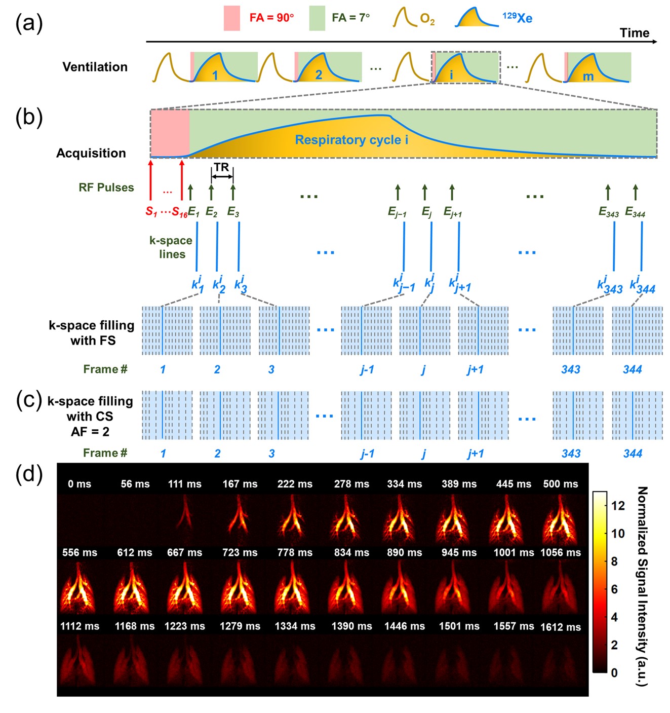
The schematic diagram of ventilation, data acquisition, and k-space line-filling strategies for dynamic ventilation imaging
Using this technology, the research team detected a significantly reduced gas MRI signal-time curve with regional variations in the lungs of rats with lipopolysaccharide (LPS)-induced lung injury (Figure 2a-f). Based on this, the research team has also developed a set of quantitative parameter systems for dynamic ventilation function, which includes parameters such as gas arrival time (Tarrival), signal peak time (Tpeak), inspiratory flow rate (Flowin), and expiratory flow rate (Flowout) (Figure 2g-i). After analysis, it was discovered that there was heterogeneity in the inspiratory and expiratory flow rates of rats with LPS-induced lung injury at the lobar level, and the ventilation rate in certain lung lobes was significantly lower than that in the control rats (P < 0.05).
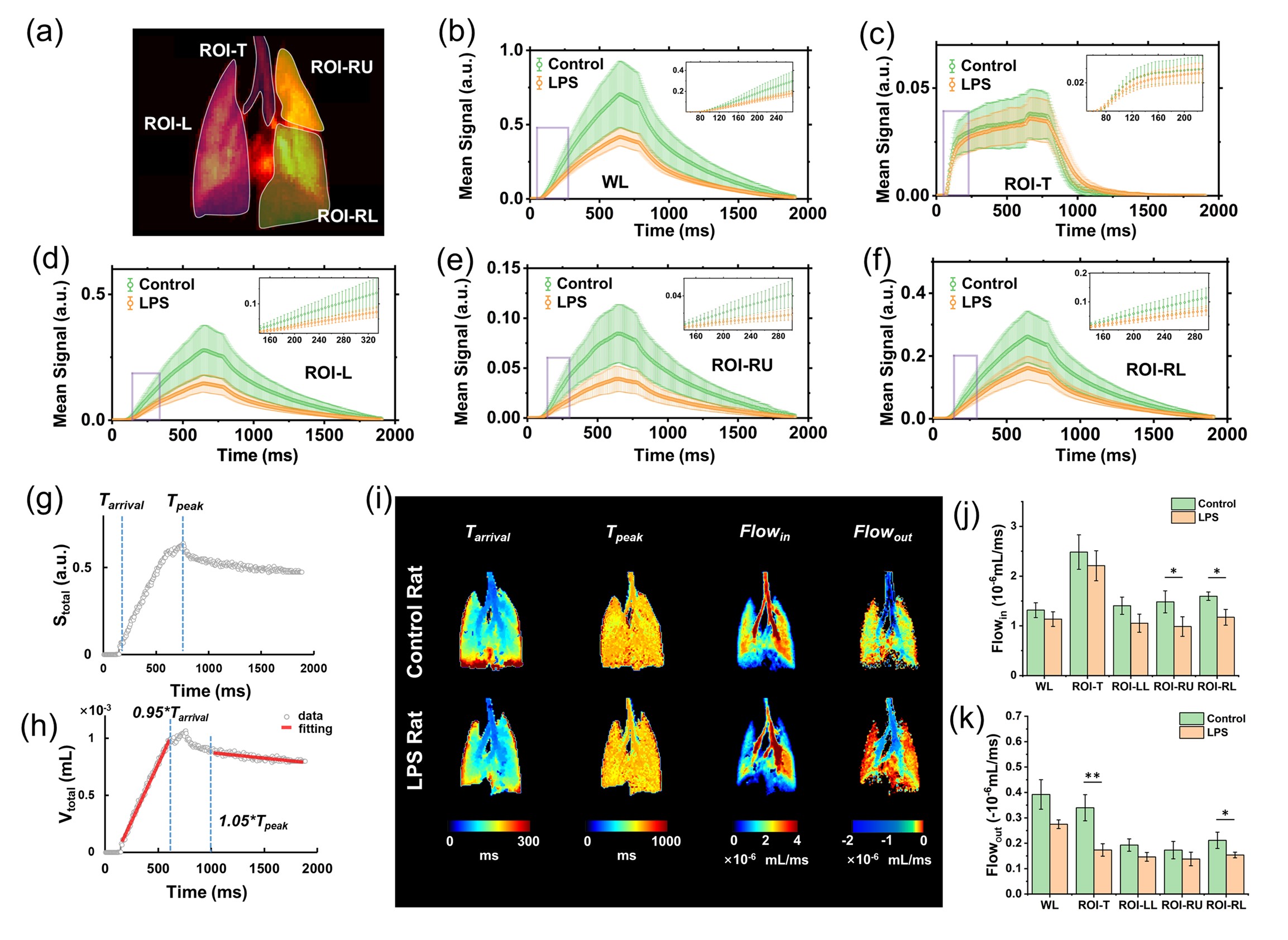
MRI signal-time curve of intrapulmonary gas; time-course diagrams of typical dynamic ventilation characteristics in the lungs, gas flow velocity distribution maps, and inter-group comparisons
The relevant research was published in Science Bulletin under the title "Dynamic ventilation functional MRI of the lung with sub-millimeter spatial resolution and millisecond temporal resolution". LI Hongchuang, a postdoctoral researcher from APM, and LI Haidong, a researcher from APM, along with FAN Li, an associate chief physician from the Second Affiliated Hospital of Naval Medical University, are the co-first authors with researcher ZHOU Xin from APM as the corresponding author.
This research was supported by projects from the National Natural Science Foundation of China (NSFC), the Chinese Academy of Sciences (CAS), and the Hubei Provincial Natural Science Foundation, among others.
Link to the article: https://doi.org/10.1016/j.scib.2025.05.014